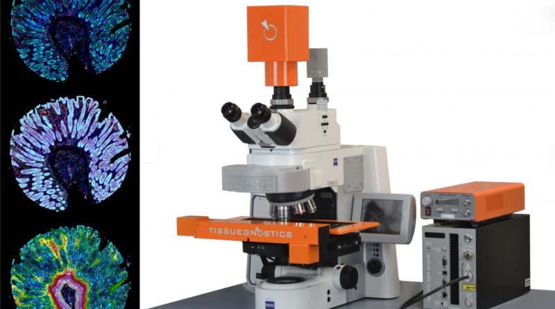
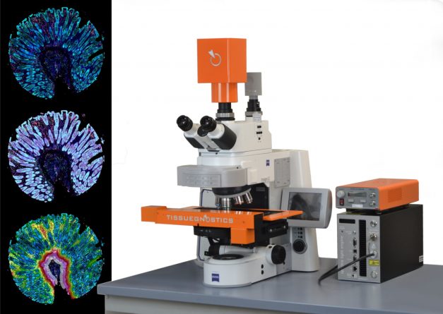
Brightfield & Multispectral Fluorescence Imaging and Spectral Unmixing

The cutting-edge multispectral tissue cytometry hardware TissueFAXS SPECTRA, developed by TissueGnostics, has the unique ability to enable multispectral imaging of IF processed samples, and spectral unmixing together with contextual tissue cytometry image analysis software (StrataQuest), in a single system. Thereby it enables high-content phenotyping in-situ.
Brightfield & Multispectral Fluorescence Imaging and Spectral Unmixing Hardware
The revolutionary SPECTRA systems are available in 8-slide standard configuration or slide loader-based configurations for automated scanning of up to 120 standard-sized slides (or 60 double-sized slides). The system supports multispectral fluorescence, widefield fluorescence and brightfield scanning. The technology includes 8 FL channels, as well as a high-quality 16-bit sCMOS camera.
The SPECTRA technology is available on any TissueFAXS system configuration, whether upright, inverted, TissueFAXS Fluo, TissueFAXS PLUS or Slide Loader-based (SL).
Multispectral imaging technology
The multispectral imaging technology by TissueGnostics is facilitated by a liquid crystal tunable filter (LCTF) and advanced spectral unmixing algorithms. LCTFs are essentially optical filters that leverage electronically controlled liquid crystal elements to transmit a selectable wavelength range of light and exclude others, for remarkable chemical imaging possibilities.
LCTFs can be tuned to specific wavelengths within the visible spectrum at high speed, making them perfect for rapid building of lambda stacks (Spectral Cubes) which are integral for spectral unmixing. Separation of multiple colours in both, brightfield and fluorescence is made possible.
Spatial and high-content phenotyping
By implementing this technology autofluorescence and channel bleed-through can be eliminated and the number of acquired IF stained markers can be dramatically increased. Besides other uses, this technology enables applications such as spatial and high-content phenotyping.
Contextual tissue cytometry analysis software for multispectral images
Unmixed channel images can then be conveniently used for follow up contextual tissue cytometry analysis as supported by the next-generation StrataQuest image cytometry solution.
At present, for drug discovery programs and research, and particularly for precision medicine, looking at multiple markers in parallel to gain a deeper understanding of the interactions of cells and cellular subpopulations, contextual tissue cytometry analysis is a key focus area of technological advancement and progress, and multispectral tissue cytometry underpins this perfectly clearing the road for even more advancements.
Advancing drug discovery and development with multispectral imaging
Multispectral imaging and spectral unmixing can support your R&D with:
- Multispectral, widefield fluorescence & brightfield whole slide scanning
- Fluorescence and Brightfield acquisition and analysis of IF/HE/IHC processed tissue sections, smears and Tissue Microarrays (TMA)
- Quantitative image analysis
- Single-cell (bio)marker quantification and high dimensional co-expression and phenotyping
- Analysis of spatial relationships between cellular subpopulations as well as metastructures
- Determination of the immune status in situ
- Detection and quantification of subcellular markers and pathogens
- Supports FISH/CISH/RNAscope imaging and analysis
TissueFAXS SPECTRA System Features
- Upgradable platform
- 8-slide stage or 120 slide loading system
- Extended depth of focus
- Compatible with standard and double-sized slides
- High powered multi-LED light engine
- High-Speed fluorescence camera option
- One click-automation
- Oil objectives
- Compact design
- Easy maintenance
- Integrated image analysis software
To learn more about how the TissueFAXS SPECTRA multispectral fluorescence & brightfield scanner can benefit your cell line development, research or drug discovery project please complete the Quick Contact form or email: office@tissuegnostics.com
You can also read more about TissueFAXS SPECTRA or request a quote here: