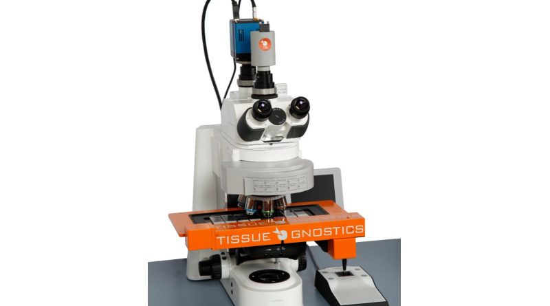
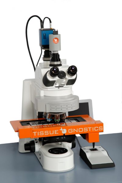
High-content Screening System

The cutting-edge TissueFAXS PLUS high-content screening system by TissueGnostics is engineered for advanced multi-channel fluorescence microscopy and exceptional high-resolution imaging to further cell research and drug discovery programs.
High-content screening system for drug discovery
High-content screening (HCS) systems are key technologies for today’s drug discovery strategies to ensure candidate compounds can fail early and cheaply in the preclinical study stage, rather than more costly later in the clinical phase.
Automated brightfield and fluorescence slide scanner
Designed to offer flexibility the TissueFAXS PLUS configuration is an upright whole slide scanning system, focusing on automation in scanning brightfield and immunofluorescence slides, cytospins, smears, and TMAs.
The microscope-based system is highly customisable and upgradable (multispectral and confocal imaging) to answer the needs of researchers over time. The TissueFAXS PLUS can be configured and modified with high-end hardware such as high powered multi-LED light engines, highly sensitive sCMOS cameras, and multiband bandpass filters for high-speed fluorescence scanning.
Powerful integrated image analysis software
The TissueFAXS PLUS comes complete with powerful integrated image analysis software for single cell quantification (HistoQuest and TissueQuest) and/or contextual image analysis with integrated AI modules (StrataQuest). The accurate and reliable image analysis software leverages rich information from the image-based screens allowing researchers to focus on those compounds that give rise to the right phenotypic changes without undesirable effects.
Accelerating drug discovery and cell research
TissueFAXS high content screening is unique in its ability to combine automated high-resolution imaging with the scientific accuracy of flow cytometry. The TissueFAXS high-content screening systems can help accelerate drug discovery, drug development and research in numerous therapeutic fields, including oncology, immunology and neurology.
The fully automated tissue cytometer can support your R&D with:
- Fluorescence & brightfield whole slide scanning of tissues, TMAs, smear and cultured cells on slides processed by immunofluorescence and immunohistochemistry
- Live cell imaging
- Quantitative image analysis
- Single-cell (bio)marker quantification and high dimensional co-expression and phenotyping
- Analysis of spatial relationships between cellular subpopulations as well as metastructures
- Determination of the immune status in situ
- Detection and quantification of subcellular markers and pathogens
- Supports FISH/CISH/RNAscope imaging and analysis
TissueFAXS PLUS High-content Screening System Features
- Upgradable platform
- 8-slide stage
- Extended depth of focus
- Compatible with oversized slides
- One click-automation
- Oil objectives
- High-Speed fluorescence camera option
- High powered multi LED light engine
- Integrated image analysis
To learn more about how the TissueFAXS PLUS platform can benefit your cell line development, research or drug discovery project please complete the Quick Contact form or email: office@tissuegnostics.com
You can also read more about TissueFAXS PLUS or request a quote here: