Spinning Disk Confocal Imaging and High-Throughput Scanning Systems

The next-gen TissueFAXS Q confocal imaging systems are highly flexible scanning platforms which offer the advantages of both laser-free ultra-fast spinning disk confocal imaging and high-throughput slide scanning.
Automated whole slide confocal imaging
Developed by TissueGnostics, a global leader in tissue-based cytometry, the state-of-the-art TissueFAXS Q platform, delivers rapid automated whole slide confocal imaging using a confocal spinning disk, paired with a high-power multi-channel LED light engine, a high-end sCMOS camera and the TissueFAXS automated scanning workflow.
Spinning disk confocal imaging for precision drug discovery
The TissueFAXS Q spinning disk confocal imaging systems are a powerful tool for rapid high-resolution cell imaging. Along with the latest generation improvements in optical design and camera technology TissueFAXS Q provides high-resolution images and optical sectioning suitable for high-end quantification analysis essential for modern precision drug discovery. Similarly to laser scanning, spinning disk confocal microscopes are capable of acquiring thin optical sections from specimens, only considerably faster, clearing the path to obtaining images at previously unattainable rates.
Flexible confocal imaging and high-throughput scanning
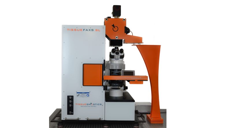
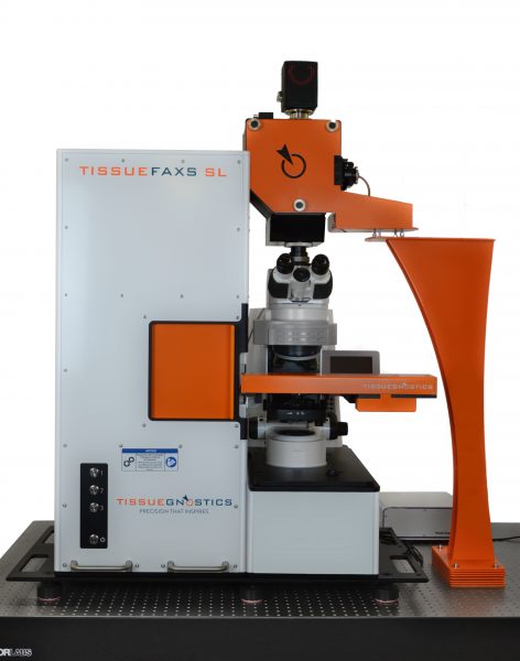
Confocal imaging systems of the TissueFAXS Q line are available in three configurations, TissueFAXS Q+, TissueFAXS iQ+ and TissueFAXS SL Q+.
The systems can have either an upright (Q+) or inverted (iQ+) design and slide loader (SL) configuration. The TissueFAXS SL Q+ is the most advanced system variation of the line.
The TissueFAXS Q+ is the upright configuration for automated high-resolution scanning using an 8 slide stage, 16bit sCMOS camera and high-power LED light engine. The system is also capable of brightfield as well as widefield fluorescence scanning.
The inverted TissueFAXS iQ+ system allows for confocal, widefield fluorescence and brightfield scanning for up to 8 slides. This includes cultured cells in well plates and petri dishes as well as live cell imaging.
The advanced ultra-fast TissueFAXS SL Q+ model is additionally equipped with a slide loader providing high-throughput scanning for up 120 slides at a time in brightfield, widefield fluorescence, and confocal fluorescence.
Through the fluid co-functionality of its cutting-edge modules, the TissueFAXS SL Q can scan a complete slice of mouse brain (73.3 mm2) with 4-channels and 13 confocal z-stacks at 20x magnification in only 1.5 hours.
The TissueFAXS Q platform also comes with the latest generation integrated image analysis software for single cell quantification, HistoQuest and TissueQuest, and/or the StrataQuest AI-assisted contextual image analysis software.
Accelerating R&D and precision drug discovery
The TissueFAXS Q platform of spinning disk confocal imaging and high-throughput screening systems can help accelerate precision drug discovery and research in numerous therapeutic fields, including oncology, immunology and neurology.
The automated confocal imaging systems can support your R&D with:
- Confocal high-throughput whole slide scanning
- Fluorescence & brightfield whole slide scanning of tissues, TMAs, smear and cultured cells on slides processed by immunofluorescence and immunohistochemistry
- Extended depth of focus and Z stacking
- Live cell imaging
- Quantitative image analysis
- Single-cell (bio)marker quantification and high dimensional co-expression and phenotyping
- Analysis of spatial relationships between cellular subpopulations as well as metastructures
- Determination of the immune status in situ
- Detection and quantification of subcellular markers and pathogens
- Supports FISH/CISH/RNAscope imaging and analysis
TissueFAXS Q Next-gen Confocal Imaging System Features
- Confocal spinning disk
- 8-slide stage or 120 slide loading system
- Brightfield scanning
- Multi-channel fluorescence scanning
- 10 filter cube positions
- 16-bit sCMOS camera
- One click-automation
- Air/Oil objectives (1x to 100x)
- High powered solid state multi LED light engine
- Slide ID scanner and barcode reader
- Integrated image analysis software
To find out more about the TissueFAXS Q platform and how it can advance your cell line development, research or drug discovery project please complete the Quick Contact form or email: office@tissuegnostics.com
You can also read more about TissueFAXS Q or request a quote here: