
Microglia Demyelination Shields CNS from Axonal Damage
A groundbreaking study by Groh et al., recently featured in Nature Communications, delves into the interplay between adaptive immunity and neurodegeneration within the central nervous system (CNS). Their research uncovers a previously unrecognized inverse correlation between demyelination and axonal damage, suggesting that microglia-facilitated demyelination may serve as a defense mechanism, shielding axons from cytotoxic T-cell-mediated destruction. Utilizing genetically modified mice with alterations in proteolipid protein (PLP) – the key constituent of myelin sheaths – the researchers identified several contributing factors to axonal deterioration, including cytoskeletal alterations in oligodendrocytes and paranodal constrictions. By employing Striatech’s OptoDrum system to assess visual acuity, the study found significant declines in the PLPmut mice, highlighting the impact of PLP defects on retinal health and axonal integrity. These discoveries provide insight into the origins of axonal spheroids, a hallmark of neurodegeneration.

PUBLICATION
Nature Communications (Oct 30, 2023) “Microglia-mediated demyelination protects against CD8+ T cell-driven axon degeneration in mice carrying PLP defects”
Groh J, Abdelwahab T, Kattimani Y, Hörner M, Loserth S, Gudi V, Adalbert R, Imdahl F, Saliba AE, Coleman M, Stangel M, Simons M, Martini R
PLP Dysregulation in Mouse Models Reveals Divergent Axonopathic Phenotypes
The study examined two distinct mouse models with modified PLP expression: one model with overexpressed PLP (PLPgt) and another with a point mutation causing defective PLP (PLPmut). Both models exhibited axonal degeneration, marked by the presence of axonal spheroids and heightened cytotoxic T-cell activity in adjacent white matter. However, their clinical profiles diverged: PLPmut mice demonstrated significant motor coordination impairment, whereas PLPgt mice did not.
Additionally, Striatech’s OptoDrum system revealed a marked decline in visual acuity in PLPmut mice but not in PLPgt counterparts. These pathological changes correlated with a significant depletion of retinal ganglion cell populations and a reduction in the thickness of the inner retinal layer. Further analysis using electron microscopy highlighted severe axonal deterioration in the optic nerve of PLPmut mice, reinforcing the idea that this model experiences heightened neuroaxonal damage.
While both PLPmut and PLPgt mice developed axonal spheroids at the nodes of Ranvier, key differences emerged. The spheroids in PLPgt mice were generally smaller and frequently exhibited demyelination, particularly as the disease advanced. Surprisingly, despite the more extensive demyelination, PLPgt mice retained greater axonal integrity in the optic nerve than PLPmut mice. This raised an intriguing question: how does increased demyelination correlate with reduced axonal degeneration?
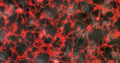
Microglial-Oligodendrocyte Crosstalk Mediates PLP-Linked Demyelination
To explore the influence of microglia in PLP-related neurodegeneration, researchers conducted single-cell RNA sequencing. They identified activated microglia (AMG) populations in both models, characterized by elevated cellular stress markers and pro-inflammatory signaling. Notably, PLPgt mice exhibited an additional microglial subtype, AMG2, which displayed heightened phagocytic activity. This finding suggested that microglia in PLPgt mice were actively clearing myelin, potentially explaining the observed increase in demyelination. But what role did this play in modulating cytotoxic T-cell responses?
Demyelination by Microglia Reduces Axonal T-Cell Damage
To determine whether microglial demyelination served a neuroprotective function, researchers carried out two critical experiments. First, they stimulated microglial activity in PLPmut mice using a cuprizone-based toxin, which resulted in enhanced demyelination. This intervention unexpectedly led to reduced axonal loss and a rise in AMG2 microglia. Second, they eliminated microglia in PLPgt mice using PLX5622, which suppressed demyelination but triggered more extensive axonal deterioration. These findings indicate that microglia play a crucial defensive role by stripping away myelin, thereby reducing the susceptibility of axons to cytotoxic T-cell targeting.
Paranodal Constriction: A Key Driver of Axonal Injury
Previous studies suggested that juxtaparanodal domains – regions adjacent to the nodes of Ranvier – serve as hotspots for axonal spheroid formation, often accumulating cellular debris. This study introduced a novel mechanism: paranodal constriction directly correlates with axonal swelling and spheroid formation. In PLPmut mice, increased paranodal constriction was linked to impaired axonal transport and the accumulation of cellular debris, potentially disrupting intracellular organelle distribution.
To confirm that paranodal constriction resulted from adaptive immune activity, the researchers suppressed adaptive immune responses, which led to reduced constriction and diminished axonal degeneration. These findings suggest that cytotoxic T-cell assaults on myelinated fibers drive paranodal narrowing, leading to impaired neuronal function.
Cytoskeletal Adaptations in Oligodendrocytes Under Immune Stress
Transcriptomic profiling of oligodendrocytes revealed elevated expression of genes associated with cell repair and cytoskeletal dynamics. Further studies identified an increase in filamentous actin (F-actin) and phosphorylated myosin light chain (pMLC) at paranodal sites, with immune-dependent amplification. When isolated oligodendrocyte progenitors were exposed to cytotoxic molecules, they exhibited similar increases in F-actin and pMLC levels, suggesting that cytoskeletal remodeling within oligodendrocytes underpins paranodal constriction.
This mechanism resembles Schwann cell-mediated axonal constriction in the peripheral nervous system, where constriction facilitates axonal fragmentation and subsequent regeneration.
Axonal Spheroids Impair Transport via Cytoskeletal Disruption
The researchers also examined whether axonal spheroids impede intracellular transport, as suggested by prior studies. Their findings confirmed that PLPmut mice exhibited significantly slower axonal transport rates, particularly in neurons with larger spheroids. This impairment was closely linked to cytoskeletal modifications. Notably, administering Fasudil, a Rho-associated kinase (ROCK) inhibitor, reduced axonal spheroid formation without altering cytotoxic T-cell activity in white matter. This suggests that Fasudil mitigates T-cell-induced actomyosin contractions at paranodal junctions, thereby preserving axonal transport efficiency.
Implications for Neurodegeneration and Future Therapeutics
This study highlights an inverse relationship between demyelination and neurodegeneration, where microglia function as protectors by selectively removing compromised myelin before cytotoxic T-cells can target axons. The findings also propose that oligodendrocytes respond to T-cell-induced stress by remodeling their cytoskeleton, leading to paranodal constriction and subsequent disruptions in axonal transport. This mechanism may represent a CNS parallel to Schwann cell-mediated axonal restructuring in peripheral nerves.
Crucially, the study identifies novel therapeutic pathways for neurodegenerative diseases. By inhibiting actomyosin contractions, Fasudil presents a potential intervention to alleviate axonal damage. Further exploration into microglial regulation and cytoskeletal modifications may yield innovative treatments for multiple sclerosis and other demyelinating disorders.
Groh et al.’s findings challenge the prevailing notion that demyelination is solely detrimental, instead highlighting a complex interplay between adaptive immunity, oligodendrocytes, and microglia. This research offers valuable insights into CNS neurodegeneration and opens new avenues for therapeutic advancements aimed at preserving neuronal integrity.
Original Source: Emilia Kawecka, Technical University of Munich, Student Assistant at Striatech
Original Paper: Groh, J., Abdelwahab, T., Kattimani, Y. et al. Microglia-mediated demyelination protects against CD8+ T cell-driven axon degeneration in mice carrying PLP defects. Nat Commun 14, 6911 (2023). https://doi.org/10.1038/s41467-023-42570-2
Related Product