
Evaluating Photoreceptor Replacement as a Viable Therapy in a Late-Stage LCA Mouse Model
Retinal degeneration represents a primary cause of irreversible blindness, with both its onset and severity differing significantly among various conditions. One of the most severe forms is Leber congenital amaurosis (LCA), a rare genetic disorder that triggers rapid and early photoreceptor deterioration, with cone cells frequently disappearing entirely by the age of three in affected individuals. Because of this early degeneration, second-order neurons, such as bipolar cells are also affected during their developmental phase, leading to abnormal synapse formation and remodeling of the inner retina. These downstream consequences pose substantial challenges for restorative therapies.
In a recently published study, Christopher Procyk and colleagues from Rachael Pearson’s laboratory at King’s College London investigated whether human cone photoreceptors (hCones), derived from pluripotent stem cells, can achieve functional integration into an LCA model exhibiting advanced degeneration. Expanding on earlier research that showed successful hCone transplantation in the less severe rd1 mouse model (1), this study examined the potential for the therapy to be effective in a late-stage context. The results show that hCone transplantation promotes structural remodeling and leads to partial recovery of visual function, even in a model with severe inner retinal disruption.

PUBLICATION
Stem Cell Reports (Apr 08, 2025) ”Human cone photoreceptor transplantation stimulates remodeling and restores function in AIPL1 model of end-stage Leber congenital amaurosis”
Procyk CA, Melati A, Ribeiro J, Liu J, Branch MJ, Delicata JD, Tariq M, Kalarygrou AA, Kapadia J, Khorsani MM, West EL, Smith AJ, Gonzalez-Cordero A, Ali RR, Pearson RA
Evaluating Transplantation in the Aipl1−/− Model of LCA
To evaluate therapeutic potential under severe degeneration conditions, researchers utilised an Aipl1−/− mice model, which is a widely attested model that mirrors the early and aggressive photoreceptor degeneration seen in LCA. These mice experience near-total cone loss by postnatal day 60, along with significant inner retinal remodeling, including synaptic disorganisation and reactive gliosis.
Upon reaching three months of age, well beyond the stage of photoreceptor degeneration, Aipl1−/− mice were given subretinal transplants of 500,000 GFP-labeled hCones in each eye. At six months (three months after the transplantation), retinas were analysed for structural integration, synapse formation, and visual function.
Profile of the Retinal Environment Prior to Transplantation
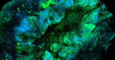
Immunohistochemical evaluation prior to the transplantation showed a complete lack of cone photoreceptors in the mid-central region of the retina. In reaction to this degeneration, bipolar and horizontal cells displayed marked dendritic retraction, with horizontal cells also exhibiting abnormal morphological features. Conversely, inner retinal neurons such as amacrine cells and retinal ganglion cells (RGCs) remained structurally preserved, indicating some degree of maintenance despite severe outer retinal degeneration.
Importantly, Müller glia showed increased expression of glial fibrillary acidic protein (GFAP), a key indicator of reactive gliosis and scarring. Synaptic markers were also substantially downregulated – RIBEYE (presynaptic) and mGluR6 (postsynaptic) were almost imperceptible in the outer retina, signifying disrupted ribbon synapse assembly. These findings confirmed the retina’s profoundly degenerative state, establishing a substantial challenge for effective therapeutic intervention.
Host Neuronal Remodeling Triggered by hCone Transplantation
Following transplantation, hCones formed multilayered clusters from 10 to 15 layers positioned adjacent to the inner nuclear layer (INL). These donor photoreceptors expressed key photoreceptor-specific proteins and demonstrated strong structural integration.
Host bipolar and horizontal cells extended their dendritic processes into the donor grafts. Notably, certain rod-specific ON-bipolar cells were observed migrating into the donor cell mass. Additionally, Müller glia infiltrated the transplant site, implying a potential functional role akin to their normal involvement in retinal support and visual pigment recycling.
Importantly, hCones upregulated RIBEYE. This upregulated co-occurred with the reappearance of mGluR6 on the dendrites of host bipolar cells, suggesting the early stages of synaptogenesis. While the full maturation of ribbon synapses remains uncertain, the presence of these markers, together with the functional data presented below, points to active glutamatergic signal transmission.
Evidence for Visual Function Recovery from Multiple Modalities
Although full-field electroretinograms (ERGs) failed to detect measurable signals in both transplanted and untreated Aipl1−/− mice, this outcome was anticipated. ERGs evaluate overall retinal activity, while the transplanted area constituted only around 10% of the total retina, comparable in size to the human macula.
Conversely, localised micro-electroretinograms (mERGs) recorded reproducible light-evoked responses within the graft region, confirming functional integration between donor and host cells. While these signals were weaker than those observed in Gnat1−/− mice (a rod-deficient, cone-only control), they were clearly distinguishable from responses in untreated Aipl1−/− mice.
Spike sorting, a key analytical method in neuroscience used to isolate and characterise the firing patterns of individual retinal ganglion cells (RGCs), showed a marked increase in light-responsive units post-transplantation, with a particular enhancement of ON responses. Notwithstanding, ON-OFF responses remained underrepresented relative to control mice, implying that some aspects of light processing remain impaired. This may indicate incomplete or maladaptive synaptic wiring between transplanted hCones and host retinal circuits.
Latency analyses demonstrated that response times in transplanted mice were comparable to those in cone-only controls under photopic conditions. This suggests that reestablished signal transmission not only occurred but did so within normal physiological timing parameters.

Stem Cell Reports, Volume 20, Issue 4, 102470. Human cone photoreceptor transplantation stimulates remodeling and restores function in AIPL1 model of end-stage Leber congenital amaurosis. Reproduced from the original publication under the Creative Commons Attribution (CC BY 4.0) license.
Partial Recovery of Light-Evoked Head Tracking Behavior
To evaluate visually guided behavior, researchers employed Striatech’s OptoDrum system, which measures optokinetic head-tracking responses to moving visual gratings. Transplanted mice exhibited a significant improvement in visual acuity compared to controls.
Significantly, 8 out of 15 mice in the transplant group consistently displayed head-tracking behavior, indicating restored visual signal transmission to the brain. Although not all animals responded, this represents a meaningful result given the severity of the disease model. The researchers suggest that response rates might be further improved with increased light intensity, as the OptoDrum system’s irradiance is limited to that of standard computer displays.
Conclusion: Functional Recovery is Possible Even in Late-Stage Degeneration Retinas
This study broadens the therapeutic scope of photoreceptor replacement by demonstrating that hCones are capable of surviving, integrating, and functioning within a retina undergoing severe degeneration. Despite the extensive remodeling and loss of native photoreceptor input, inner retinal neurons exhibit enough plasticity to form new synaptic connections with transplanted cells.
Although it has yet to be confirmed whether these synapses are fully canonical ribbon synapses, the observed glutamatergic signaling and behavioral improvements support the presence of meaningful functional recovery. Future investigations may optimise stimulus protocols, such as varying light intensities or employing naturalistic lighting, to more accurately assess ON-OFF responses and other complex aspects of visual processing.
By establishing the efficacy of hCone transplantation in both the less severe rd1 model and the advanced Aipl1−/− model, this work provides strong evidence for its broader translational relevance. Even in cases previously considered beyond therapeutic reach, photoreceptor transplantation could represent a promising strategy for restoring vision.
Original Source: Emilia Kawecka, Technical University of Munich, Student Assistant at Striatech
Original Paper: Christopher A. Procyk et al., Human cone photoreceptor transplantation stimulates remodeling and restores function in AIPL1 model of end-stage Leber congenital amaurosis, Stem Cell Reports, 2025, Volume 20, Issue 4, 102470. DOI: 10.1016/j.stemcr.2025.102470 >>
Citation: 1. “Restoration of visual function in advanced disease after transplantation of purified human pluripotent stem cell-derived cone photoreceptors” Ribeiro, Joana et al., Cell Reports, 2021.
Related Product