Digital Whole Slide Imaging System
The MMI CellScan is a cutting-edge digital whole slide imaging system that transforms any sample into high-resolution digital data in 5D.
Compatible with almost all research microscopes and imaging modes, it opens infinite digital imaging possibilities. Researchers benefit from both quantitative and qualitative assessment of tissue morphology and pathology. Additionally, the data obtained can be integrated with other omics platforms. This flexible digital whole slide scanner is particularly well-suited for applications such as spatial transcriptomics, tumor heterogeneity, liquid biopsy, rare and single-cell isolation, digital pathology, and molecular pathology.
Digital Whole Slide Imaging System
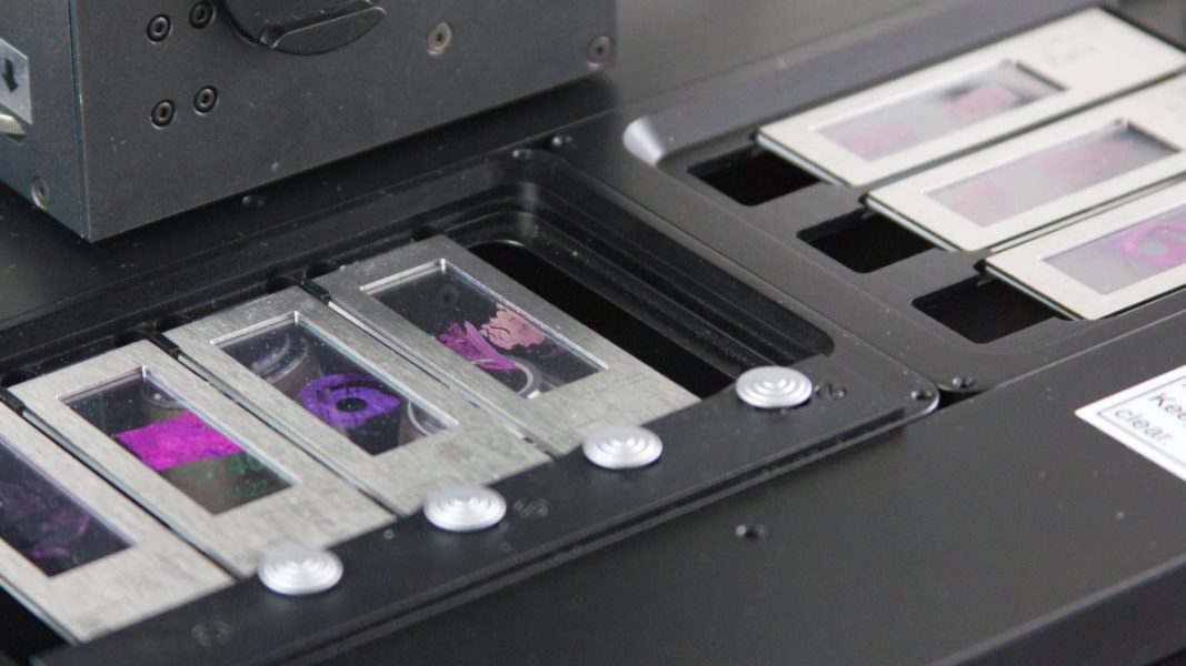
The CellScan offers unparalleled flexibility to support all image modes and sample types. By seamlessly capturing full-resolution digital slides in 5D, including time-lapse, z-stack, XY scan, and multi-color fluorescence imaging, the CellScan offers unparalleled insight into the intricate world of cellular structures and dynamics. Its support for multiple image modes allows researchers to capture dynamic changes over time, navigate sample depths, and analyze multi-fluorescence imaging with high fidelity. It scans a 15 mm x 15 mm section in less than 60 seconds and a full 75 mm x 25 mm slide in less than 7 minutes (20x objective), allowing for rapid data acquisition without compromising on image quality or resolution. From conventional glass slides to petri dishes with living cells, or membrane slides designed for laser microdissection, the CellScan accommodates a myriad of sample formats. This advanced compatibility with different experimental setups in diverse research fields, including digital pathology, cell biology, and oncology plays an instrumental role in defining optimal therapeutic strategies.
Versatile and Flexible High-Resolution Scanning System
Built on a modular design framework, the CellScan boasts compatibility with close to all research microscopes to ensure optimal performance for research or pathology services. This exceptionally flexible whole slide imaging system seamlessly integrates with all other MMI devices, such as the CellCut for laser microdissection, the CellEctor for selective isolation of rare and single cells, and the CellDetector for automated cell and tissue identification. This advanced modularity enhances its adaptability, allowing researchers to tailor their imaging setup to specific research needs facilitating a comprehensive approach to data acquisition and analysis. When combined with the MMI CellCut, CellEctor, and CellDetector, the system opens up new applications, including single-cell isolation and automated cell analysis.
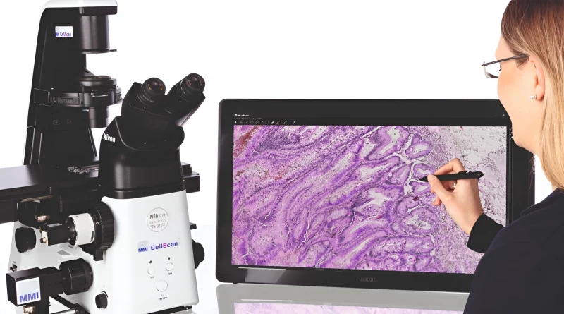
Whole Slide Imaging Software Accelerating Collaborative Research
MMI’s whole slide imaging software accelerates collaborative research and remote work, offering unlimited licenses for seamless teamwork. With instant loading of BigTIFF whole slide images, researchers can access samples from anywhere, annotate them effortlessly, and perform precise measurements, even enabling laser microdissection with the CellCut.
The software’s advanced stitching function ensures perfect image alignment, providing unparalleled image harmony and clarity, particularly beneficial for samples with complex structures or irregularities. By leveraging precise auto-focusing the reliability and reproducibility of analysis are enhanced further, especially for samples exhibiting waviness along the z and focus directions.
Whole Slide Live-Cell Imaging
With the whole slide scanner CellScan in combination with a stage-top incubator researchers are empowered to create digital slides and identify regions of interest from live images. Live cell imaging with the CellScan enables researchers to capture dynamic changes in cellular morphology and behaviour with exceptional accuracy. By facilitating time-lapse microscopy on living cells, the digital whole slide imaging system empowers researchers to study complex biological processes in real-time, shedding light on fundamental mechanisms underlying cellular function and disease pathology.
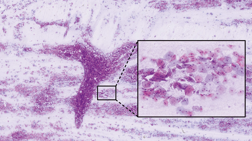
Multi-Color Fluorescence Imaging
With its ability to capture and compile multi-color fluorescence images, the whole slide imager enables researchers to investigate molecular interactions and signalling pathways with unprecedented detail. By unravelling the complexities of biological systems at the molecular level, CellScan provides insights that are crucial for advancing the understanding of disease mechanisms and therapeutic interventions.

Features:
- High-resolution digital data in 5D
- Time-lapse, z-stack, XY scan, and multi-color fluorescence imaging
- Compatible with any sample type: glass slides, petri dishes, membrane slides etc.
- Perfect image alignment & auto-focus
- Modular design for different experimental setups
- Whole Slide Live-Cell Imaging
- Collaboration & Remote Work Software
- Compatible with close to all microscopes
- Compatible with all other MMI systems